Demystifying Atrial Septal Defect; Navigating Symptoms and Treatment Options
Introduction
Congenital diseases are conditions present at the time of birth, often termed as defects that could be structural or functional. Structural defects affect the physical development of the body parts like the limbs, brain, or heart. Functional defects affect an individual's intellectual ability and are related to how the nervous system, cells, and metabolism function. These abnormalities are prenatal and are said to occur when the fetus fails to develop normally. The deaths due to congenital diseases have been reportedly a matter of concern, with nearly 240000 newborn babies estimated to die due to these diseases. Some of the most common of these abnormalities are heart defects, neurological abnormalities, and down syndrome.
Heart defects may or may not be severe at birth, with no symptoms noticed until the child attains adulthood. Some may not require any treatment, while some may prove fatal if not addressed in a timely manner. The good news is that these congenital heart diseases are no longer untreatable. A timely and effective treatment can be lifesaving, providing a better quality of life.
The present blog discusses one of the congenital heart defects, known as Atrial Septal Defect, that affects the atrial septum (the wall) that divides the atria (the upper chambers of the heart) and, thereby, the functioning of the heart.
What is an Atrial Septal Defect?
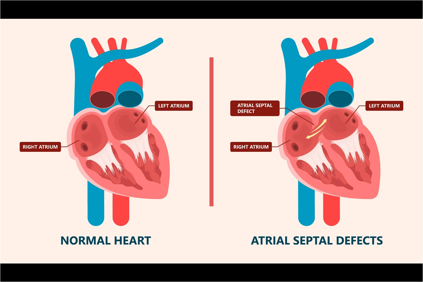
An Atrial Septal Defect (ASD) is a congenital heart disease wherein a patient is born with a hole in the atrial wall that separates the right and the left atria. The abnormality is caused during pregnancy when the baby's heart develops. Often, openings in the septum could eventually close, but if one of the openings/holes does not close, it leads to ASD. The hole in the septum varies in size. There is a disruption in the blood flow, and the disruption depends on the size of the hole. The left atrium carries oxygen-rich blood after it passes through the lungs. In the case of ASD, the blood flows from the left to the right atrium, carrying oxygen -rich blood that goes to the lungs for processing. With the backflow of blood from the left atrium, the right atrium has excess blood that has to pass through the lungs. Over a period of time, this excess amount of blood damages the lungs.
Types of ASD
- Primum ASD- the ASD is in the lower part of the septal wall and may lead to other heart abnormalities like mitral or tricuspid valve defects, Down Syndrome, or atrioventricular septal defect.
Secundum ASD- ASD is in the middle of the septal wall and is the most common of the ASDs in babies. - Sinus venosus ASD- is caused by a hole in the lower or upper back of the atrial septum. It leads to defects in the right pulmonary veins.
- Unroofed Coronary Sinus- the condition is the rarest form of ASD with no wall or incomplete wall between the coronary sinus (veins connected to the heart) and the left atrium. The condition often causes other heart defects.
Causes of ASD
A small hole in the septum may not be a serious condition so long as it does not affect the functioning of the lungs or cause damage to the right atrium or blood vessels. A child with a small hole may not experience any symptoms. Sometimes, the symptoms are experienced in adulthood or in cases with a large hole that affects blood flow with excess load on the right atrium lungs and the blood vessels. In such cases, the following symptoms may be evident.
- Shortness of breath with little activity or exercise
- Swelling on legs, feet, belly area
- Irregular or skipped heartbeats
- Heart murmur
- Frequent lungs infections
- Difficulty in breathing
Diagnosis of ASD
ASD may be diagnosed during pregnancy using prenatal tests or in cases of symptoms of ASD evident in individuals any time after birth. The healthcare professional will diagnose the condition by performing a physical examination and examining the patient's medical history. They may conduct various diagnostic tests like –
- Electrocardiography to detect any irregularity in the electrical activity of the heart
- Chest X-rays to check any damage to the atria, lungs, or blood vessels
- Transthoracic Echocardiography to detect the irregular blood flow from the left to the right atrium
- Transesophageal Echocardiography shows the location and size of ASD, the heart valves, and the damage, if any, caused.
- Intracardiac Echocardiography is used during percutaneous procedures. A tiny camera is guided to the heart to detect ASD and blood flow.
Treatment of ASD
Once diagnosed, ASD may or may not require medical intervention. In cases of a small hole, there may not be any need for surgery or percutaneous intervention. However, if the condition is severe with a large hole disrupting the blood flow that has damaged or has the potential to damage the lungs, right atrium, or blood vessels, the healthcare professional would recommend a medical intervention. This could be either non-surgical or surgical, depending on the need and circumstances of each case.
- Non-Surgical Treatment for ASD- The non-surgical treatment is a percutaneous procedure using a catheter with a septal occluder (a device) attached. Before the procedure, the location and size of the ASD and heart pressure are checked using catheterization. The catheter is guided to the heart through a vein in the groin. Once the catheter reaches the site, the septal occluder is released, and the hole is closed. The catheter is then removed. With time, tissue grows around the occluder, and it becomes part of the body.
- Surgical Treatment- In severe cases, surgery becomes necessary. The surgery may involve using the patient's own pericardium (membrane around the heart) as a tissue patch to close the hole or suturing the hole without the use of any patch. The surgery may be a minimally invasive procedure or robotic surgery, as the patients or their caregivers may opt for.
FLORET ASD from MERIL
Floret ASD Occluder is a percutaneous, transcatheter, atrial septal defect closure device. It is designed to close ASD in the secundum position or patients who have undergone a fenestrated Fontan procedure and now require closure of the fenestration. The device is low profile and well-shaped. The nitinol wires are braided, and the device retains its original shape post-deployment due to the sharp memory of the nitinol. The three polyester fabric inserts help close the duct and facilitate tissue growth over the occluder.
Conclusion
ASD, in most cases, is treatable and may not require any surgical or non-surgical treatment. It is a congenital heart disease, and the cause of ASD is unknown. However, the birthing parent needs to take care of the risk factors contributing to the condition. The mother must avoid smoking, alcohol consumption, and certain medications when planning a pregnancy and after that. A healthy lifestyle with proper prenatal care helps ensure a healthy and safe childbirth. Regular check-ups and follow-ups after childbirth are equally important and required if there are any potential chances of ASD in the newborn baby.
References
- https://my.clevelandclinic.org/health/diseases/11622-atrial-septal-defect-asd
- https://www.cdc.gov/ncbddd/heartdefects/atrialseptaldefect.html#:~:text=An%20atrial%20septal%20defect%20is,own%20or%20may%20require%20surgery.
- https://www.mayoclinic.org/diseases-conditions/atrial-septal-defect/symptoms-causes/syc-20369715
- https://www.heart.org/en/health-topics/congenital-heart-defects/about-congenital-heart-defects/atrial-septal-defect-asd